Antibodies are invaluable tools in diagnostics and research. They enable the detection, isolation, and visualization of their target antigens across a range of experimental techniques such as western blotting, immunocytochemistry, immunohistochemistry, immunoprecipitation, and ELISA. While antigens are often proteins, antibodies can also target chemical modifications or other small structures.
Despite their widespread use, antibodies have recently been criticized due to concerns about cross-reactivity and variability. These concerns are sometimes referred to as the ‘reproducibility crisis’ or the ‘antibody crisis’. At SYSY Antibodies, we are committed to addressing these challenges by thoroughly characterizing and validating our antibodies to ensure the highest standards of quality and reliability. With in-house production, we maintain full control over batch testing and quality control.
The performance of an antibody depends primarily on its affinity and specificity:
Different experimental applications require different antibody properties due to the different antigen presentation:
As a result, an antibody that proves successful in one application may fail in another. Validation must therefore be carried out separately for each intended use.
This control helps to detect background signals from secondary reagents, but does not provide any information about the quality of the primary antibody.
Observation of signals consistent with the reported molecular weights, tissue distributions, or staining patterns can aid validation. However, molecular weight alone cannot confirm specificity, as multiple proteins with similar electrophoretic mobility may be present. Literature data may also be incomplete or inaccurate.
Transfection of cells to overexpress the target antigen helps to assess specificity and sensitivity. The target protein or fragment is often fused with a reporter gene, which allows differentiation between false positive and true signals. Clean signals of expected size or localization in tissue or lysate confirm specificity. However, artificially high protein levels may not reflect physiological conditions.
Figure 1: Indirect immunostaining of formalin fixed paraffin embedded (FFPE) HEK cells transfected with mammalian expression plasmids coding for full-length ALFA- or GFP tagged nucleocapsid proteins of diverse coronavirus strains using the monoclonal anti-SARS-CoV-2 nucleocapsid antibody clone #4A8 (cat. no. HS-452 011, dilution 1:250, DAB, brown). Nuclei have been visualized by hematoxylin staining (blue). The anti-SARS-CoV-2 nucleocapsid antibody clone #4A8 is specific for the SARS-CoV-2 nucleocapsid protein and does not bind to nucleocapsid proteins of other coronavirus strains.
By blocking antibodies with their immunogen, it is possible to determine whether a signal corresponds to the antigen. If the signal disappears, specificity is likely, although cross-reactivity with similar epitopes remains possible.
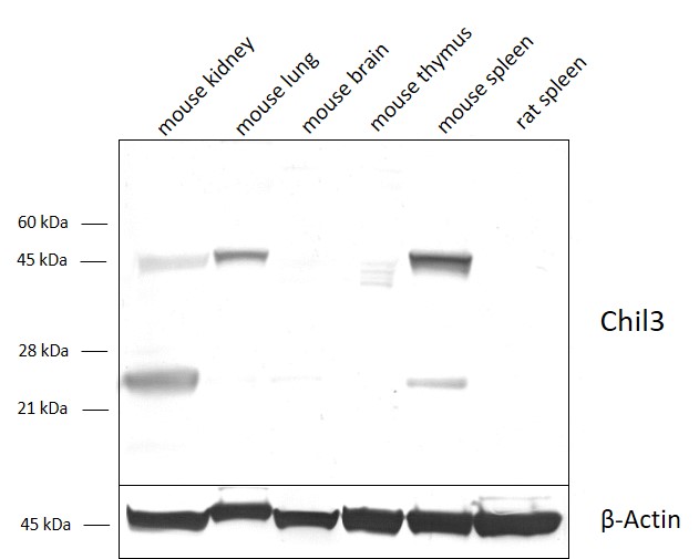
Figure 2: Detection of Chil3/YM1 with polyclonal guinea pig antibody cat. no. 442 004 in mouse spleen homogenate. Lane 1: Chil3/YM1 antibody (dilution, 1:1000). Lane 2: Chil3/YM1 antibody (dilution, 1:1000) blocked with 5 µg immunogen.
KO or KD models are the gold standard for antibody validation. Loss of signal in KO or lower signal intensity in KD tissue compared to wild type confirms specificity. However, KO or KD validation in one application (e.g. western blotting) is no guarantee of performance in other applications (e.g. immunostaining). Furthermore, although KD systems reduce the concentrations of target protein, interpretation can be difficult if KD efficiency is low.
Currently, a large percentage of our antibodies are validated using KO or KD models. We are constantly working to increase this number. If you have access to a KO model for one of our antibodies that has not yet been validated, we encourage you to contact us at marketing (at) sysy.com.
Figure 3: Detection of Galectin-3 in Galectin-3 WT and KO neuronal retina lysates with rat monoclonal anti-Galectin-3 antibody (cat. no. HS-477 017, dilution 1:500).
Courtesy: Silvia C. Finnemann, PhD, Department of Biological Sciences, Fordham University
Figure 4: Detection of HSP90 alpha and beta with rabbit polyclonal anti-HSP90 alpha (cat. no. 380 003) and polyclonal rabbit anti-HSP90 beta (cat. no. 380 103) in either HSP90 alpha siRNA KD T-RExTM-HeLa cells or HSP90 beta siRNA KD T-RExTM-HeLa cells.
Courtesy: Silvia Dalba and Prof. Stephen High, Faculty of Life Science, University of Manchester
IP followed by MS analysis is a powerful method for assessing antibody specificity in native state applications, e.g. IP or ELISA. However, these results may not correlate with specificity in denaturing methods such as western blotting.
The use of multiple antibodies against different epitopes of the same target protein can provide clear evidence of specificity. Consistent staining patterns reduce the likelihood of off-target binding.
Western Blot (WB)
In standard western blot methods, denatured protein samples are separated by their molecular weight using SDS-PAGE and transferred to a membrane. Western blotting provides information on molecular weight and insights into differential expression in different tissues or subcellular fractions such as membranes, cytosol or different organelles. However, additional background data is required to confirm the results.
Immunocytochemistry (ICC)
The ICC provides information on the subcellular localization of the target protein, but does not provide molecular weight data. Validation can be improved by co-staining with established markers. The use of different cell systems that have different expression levels or patterns of the target protein can be very helpful in analyzing specificity.
Figure 6: Indirect immunostaining of PFA fixed rat hippocampus mixed cultures with guinea pig anti-MLC1 antibody (cat. no. 525 005, dilution 1:1000, red), mouse anti-GFAP antibody (cat. no. 173 011, dilution 1:1000, white) and rabbit anti-S100B (cat. no. 287 003, dilution 1:1000, dark blue). Co-staining with the astrocyte markers GFAP and S100B shows the specific localization of MLC1 in astrocytes. Nuclei have been visualized by DAPI staining (turquoise).
Immunohistochemistry (IHC)
Evaluation of tissue specificity and sensitivity is a critical step in antibody validation to ensure reliable and accurate detection of target proteins in tissue-based experiments. Antibody testing can be performed with fresh frozen (FF), paraformaldehyde (PFA)-fixed or formalin fixed paraffin embedded (FFPE) tissue. Glyoxal fixation is an alternative tissue preservation method to formalin fixation that has gained attention due to its unique advantages over traditional formalin fixation. Each tissue preparation method offers unique benefits and challenges, so it is important to consider the specific application when planning validation experiments.
SYSY Antibodies has established a comprehensive tissue panel that includes fresh frozen, glyoxal-fixed, PFA-fixed or FFPE tissues from mouse and rat and has access to human FFPE samples from different organs. To ensure high tissue specificity and sensitivity, more and more of our antibodies are validated on both, positive and negative tissues, as well as on tissues with high and low target expression. Co-staining with known markers and comparing the observed staining patterns with in situ hybridization data or other literature data allows a better assessment of antibody specificity. This ensures that our antibodies guarantee minimal staining artifacts, a high signal-to-noise ratio, and reliable detection of the target protein in different experimental contexts.
Figure 7: Indirect immunostaining of PFA-fixed mouse lung sections of a wild type (A) or a LAMP3 knock-out mouse (B) with guinea pig anti-LAMP3 antibody (cat. no. 391 005, dilution 1:500, red). Nuclei have been visualized by DAPI staining (blue).
Courtesy: Knock-out mouse lung tissue was kindly provided by Prof. Dr. Markus Damme, Christian-Albrechts-University of Kiel
Figure 8: Immunohistochemical staining of formalin fixed paraffin embedded (FFPE) sections of (A) a cytokeratin7 (CK7)-positive human pancreatic adenocarcinoma and (B) a CK7-negative human squamous cell lung cancer engrafted into immunocompromised mice (xenografts) with rabbit anti-Cytokeratin7 (cat. no. HS-454 008, dilution 1:1000, DAB, brown). This antibody is fully human-specific and does not stain murine CK7 positive cells. Nuclei have been counterstained with hematoxylin (blue).
IP / ELISA
In IP and ELISA, the antibody must recognize native antigens. However, as the protein complexes in the analyte may still be intact, this can lead to masking of the antibody epitopes. The specificity results of these methods may not be consistent with denaturing approaches such as WB.
When KO / KD models are not available, collecting as much validation data as possible provides greater confidence in antibody specificity. Combining multiple validation methods tailored to each application helps to mitigate reproducibility and specificity issues and ultimately increases the reliability of antibody-based research.
At SYSY Antibodies, we provide detailed information on how each of our antibodies has been tested and the applications in which they have performed exceptionally well, ensuring that our customers can rely on them for their experiments.
 HEK cells transfected with mammalian expression plasmids coding for full-length ALFA- or GFP tagged nucleocapsid proteins of diverse coronavirus strains using the monoclonal anti-SARS-CoV-2 nucleocapsid antibody clone #4A8")
. Lane 2: Chil3/YM1 antibody (dilution, 1:1000) blocked with 5 µg immunogen.")

 and polyclonal rabbit anti-HSP90 beta (cat. no. 380 103) in either HSP90 alpha siRNA KD T-RExTM-HeLa cells or HSP90 beta siRNA KD T-RExTM-HeLa cells.")
, mouse anti-GFAP antibody (cat. no. 173 011, dilution 1:1000, white) and rabbit anti-S100B (cat. no. 287 003, dilution 1:1000, dark blue).")
 or a LAMP3 knock-out mouse (B) with guinea pig anti-LAMP3 antibody (cat. no. 391 005, dilution 1:500, red). Nuclei have been visualized by DAPI staining (blue).")
 sections of (A) a cytokeratin7 (CK7)-positive human pancreatic adenocarcinoma and (B) a CK7-negative human squamous cell lung cancer engrafted into immunocompromised mice (xenografts) with rabbit anti-Cytokeratin7")