Cat. No.: 171 128
Amount: 50 µg
Price:
$420.00
|
|
|
|
| Cat. No. 171 128 |
50 µg purified recombinant IgG, lyophilized. Albumin and azide were added for stabilization. For reconstitution add 50 µl H2O to get a 1mg/ml solution in PBS. Then aliquot and store at -20°C to -80°C until use. Antibodies should be stored at +4°C when still lyophilized. Do not freeze! |
| Applications | |
| Clone | RbN52 |
| Subtype | IgG1 (κ light chain) |
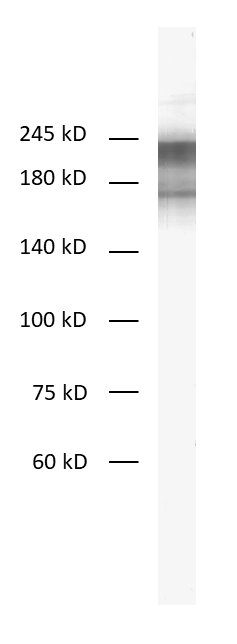
| Immunogen | Full length purified pig Neurofilament H (UniProt Id: F1RFH3) |
| Reactivity |
Reacts with: mouse (P19246), rat (P16884), pig (F1RFH3), ape, human (P12036). Other species not tested yet. |
| Specificity | Detects phosphorylated and unphosphorylated Neurofilament H |
| Remarks |
This antibody is a chimeric antibody based on the monoclonal mouse antibody N52. The constant regions of the heavy and light chains have been replaced with rabbit specific sequences. The antibody can therefore be used with standard anti-rabbit secondary reagents. The antibody has been expressed in mammalian cells. |
| Data sheet | 171_128.pdf |
|
|
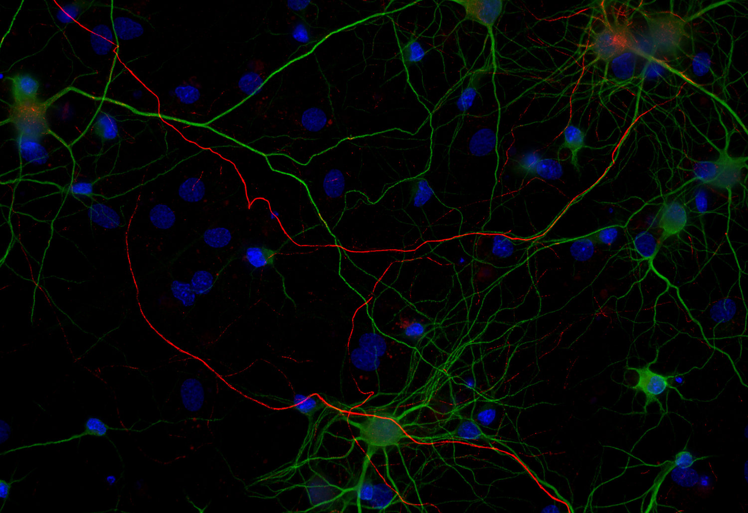
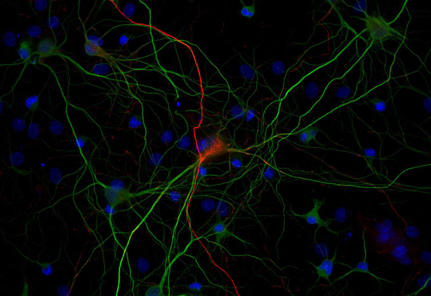
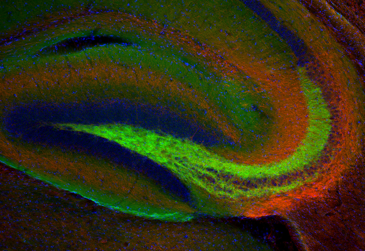
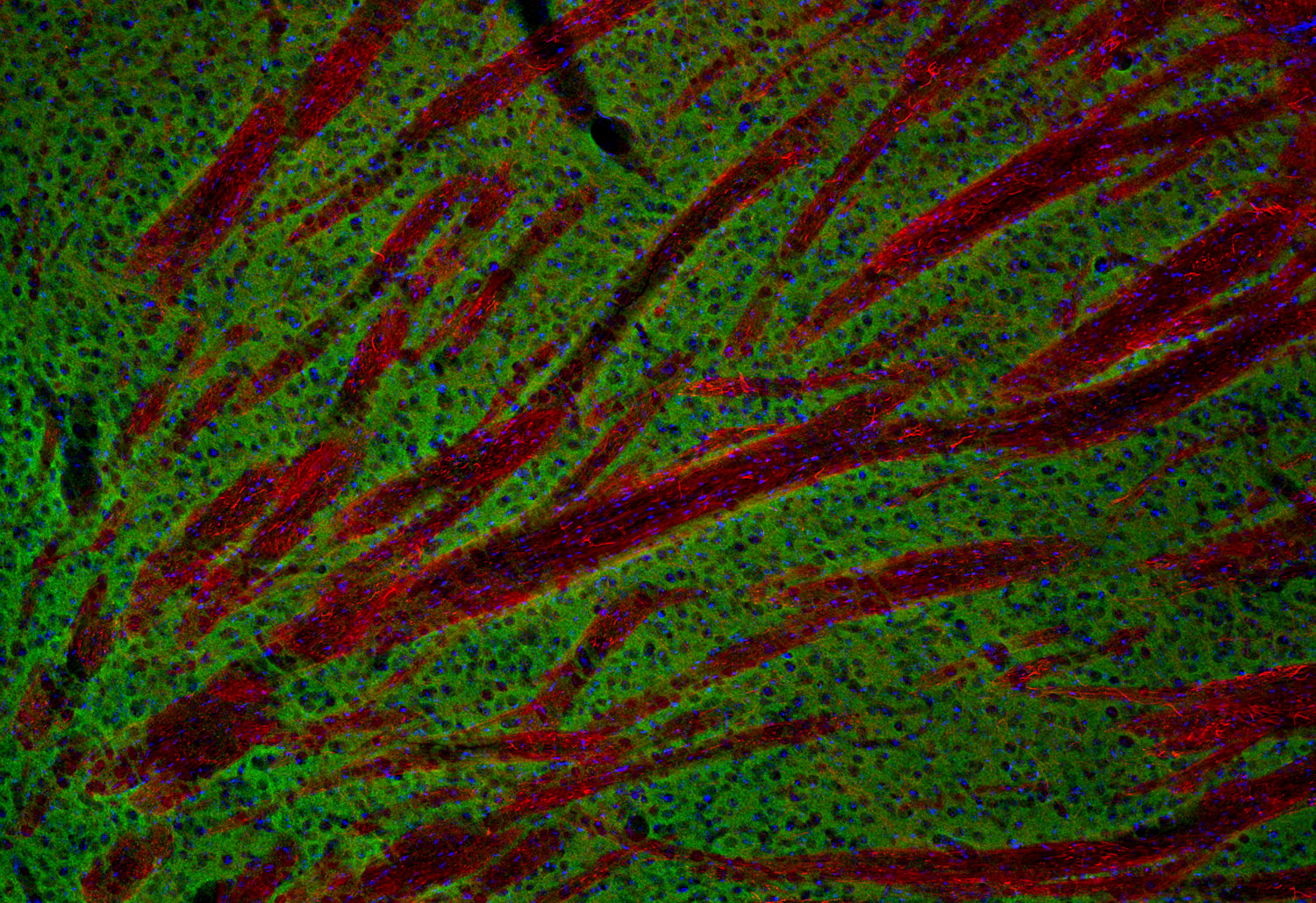
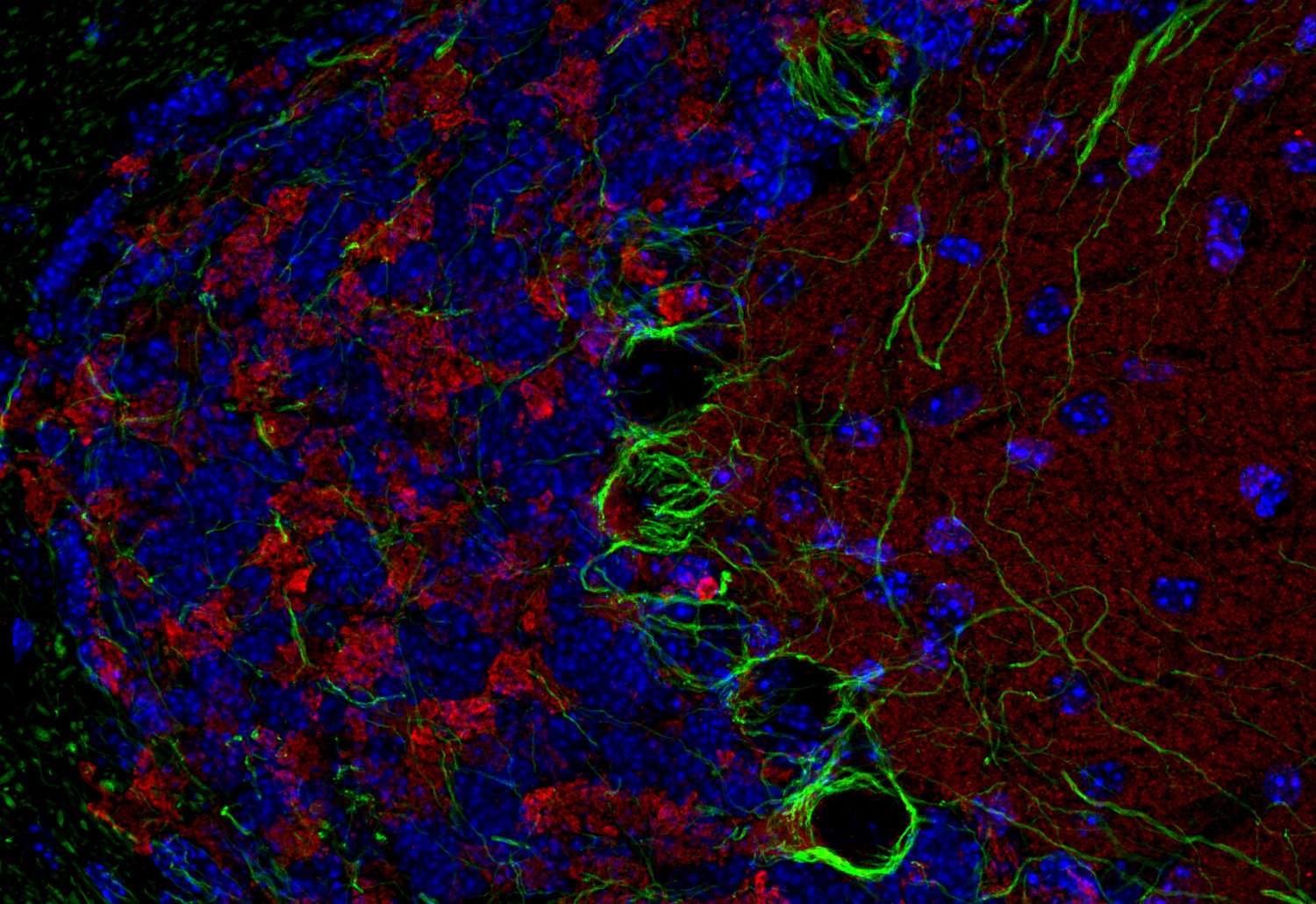
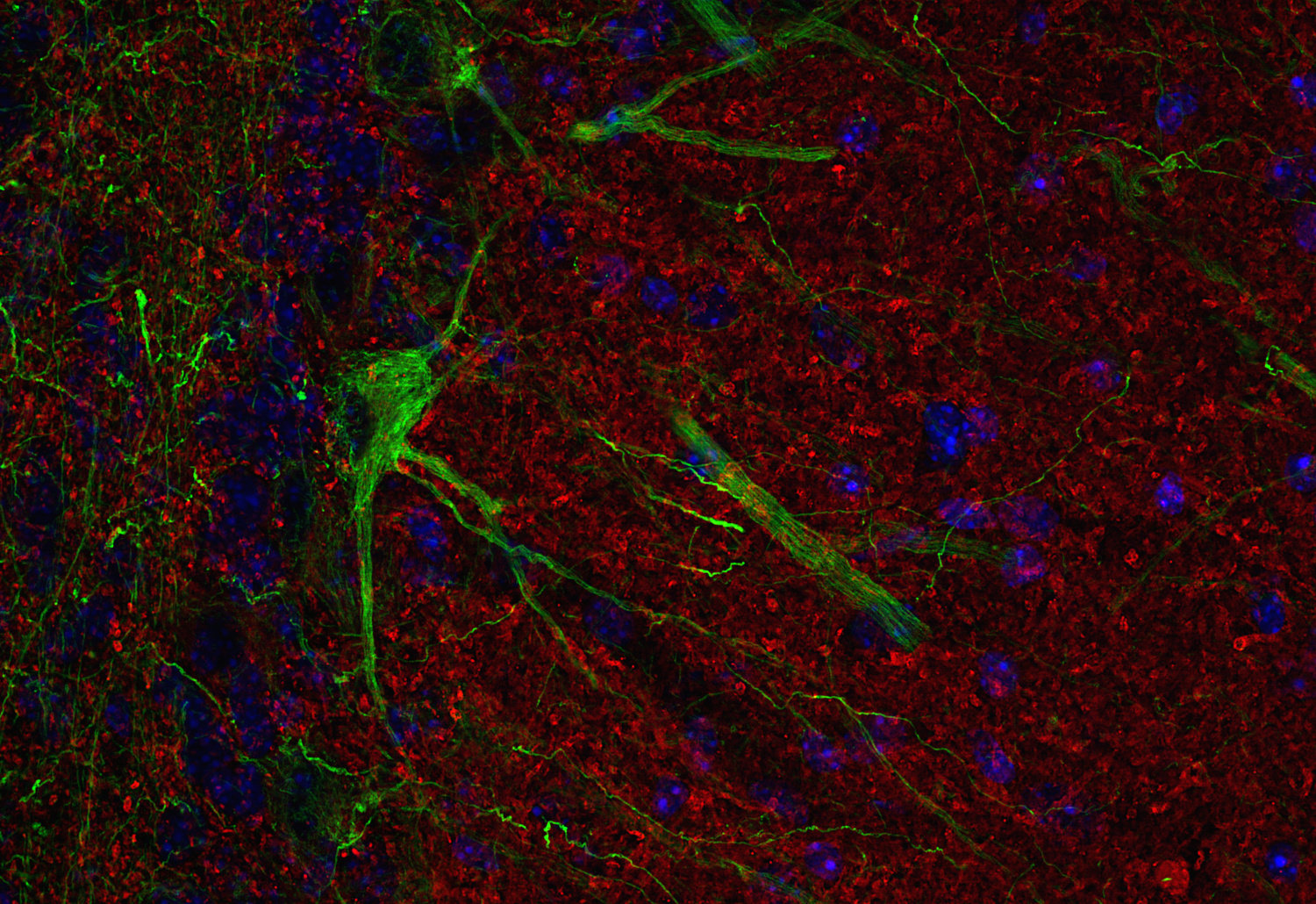
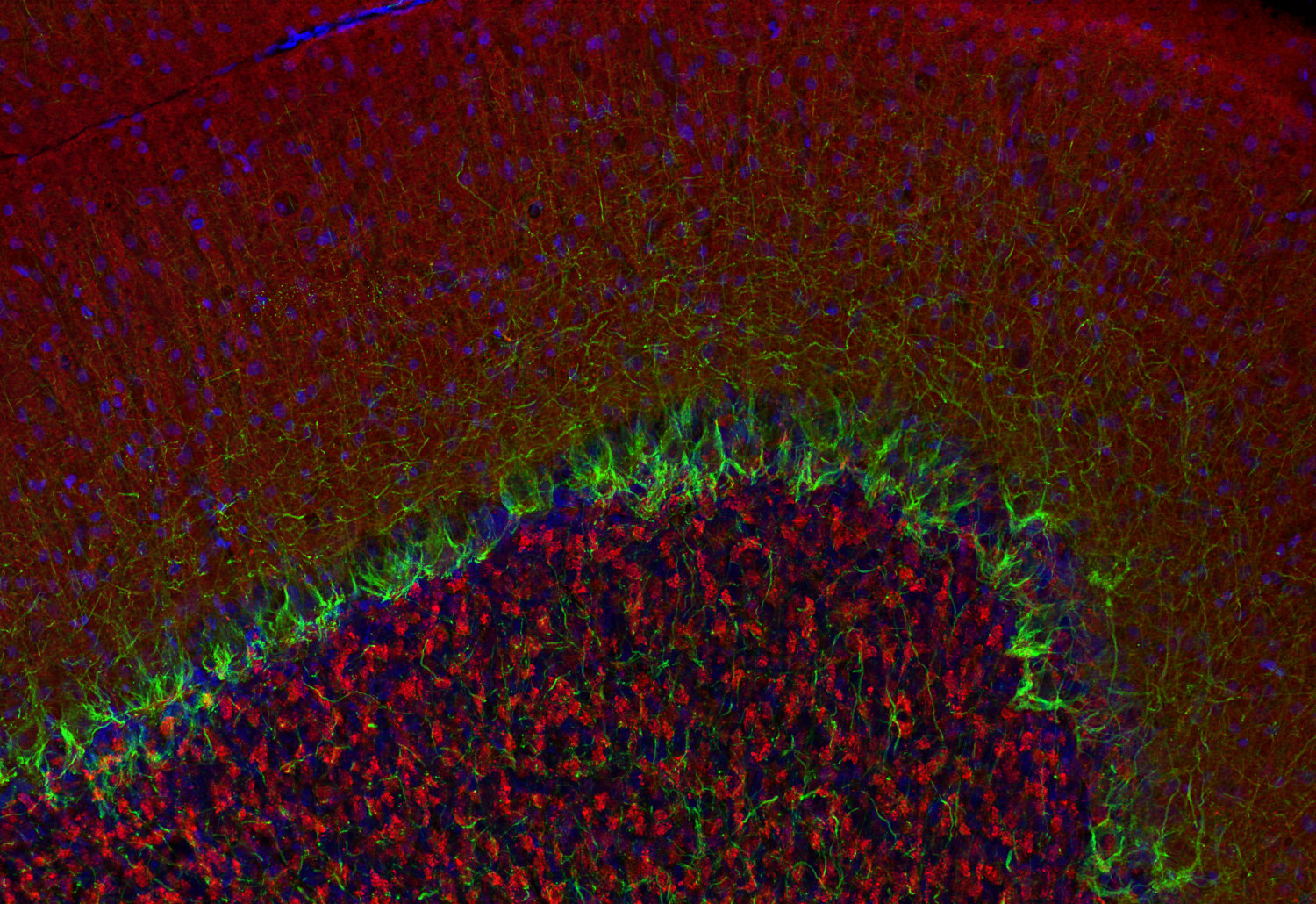
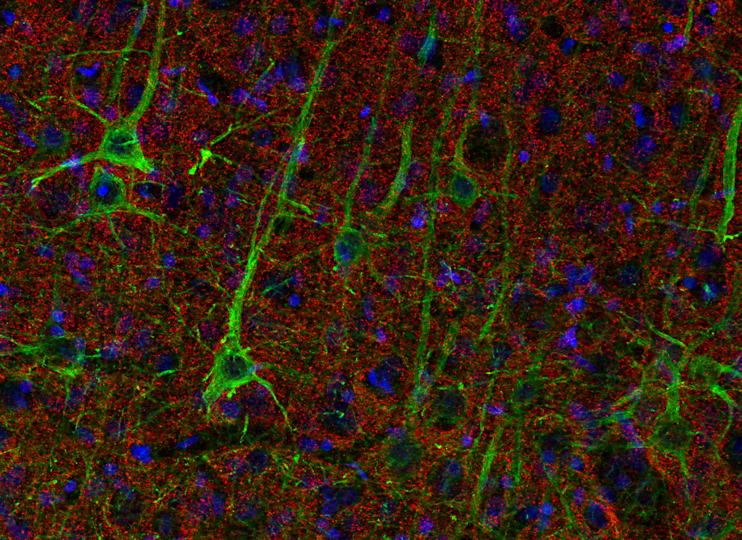
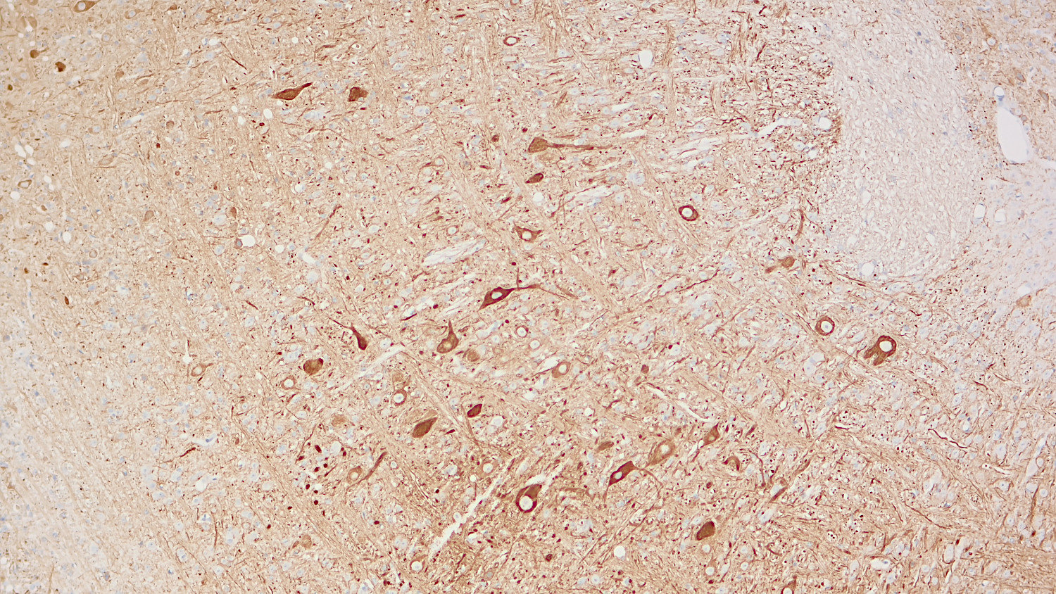

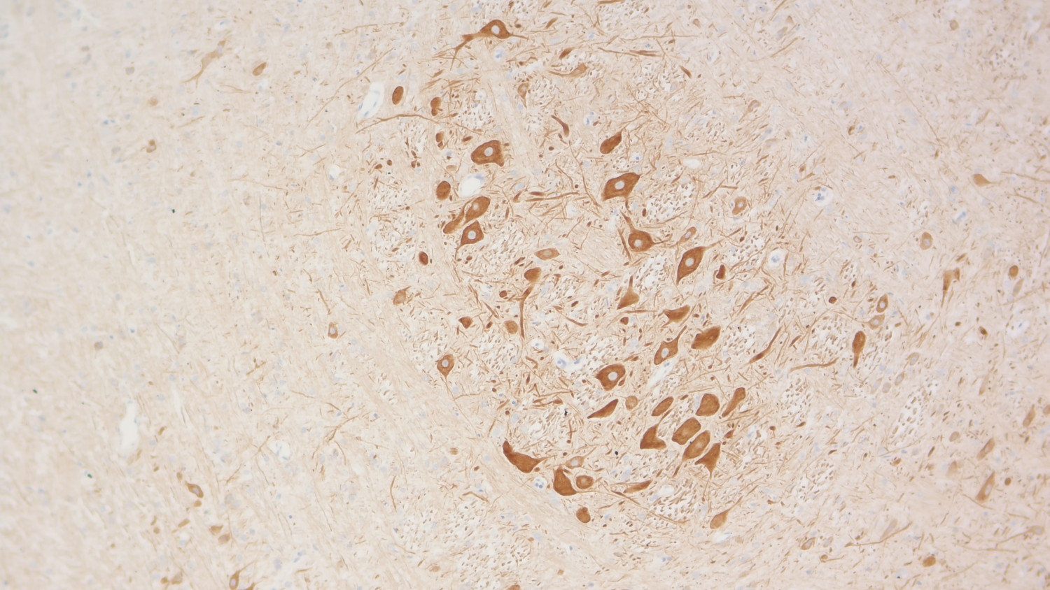
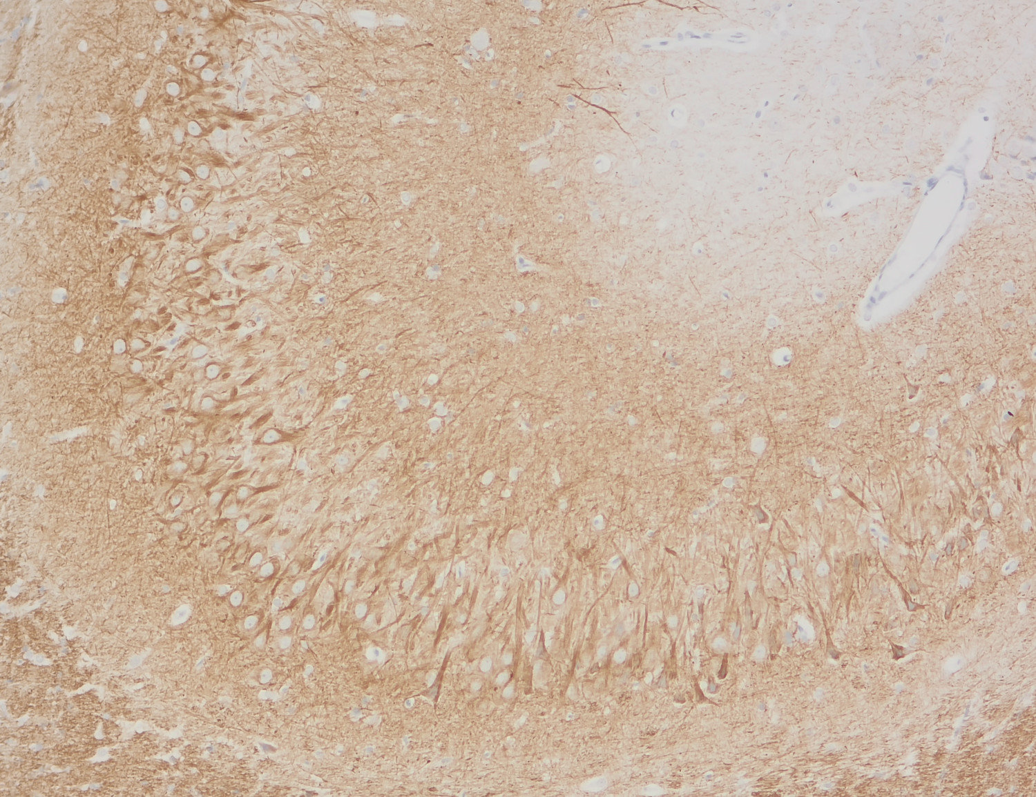
Neurofilaments (NFs) are intermediate filaments essential for providing structural support to neurons, particularly within axons. They play a crucial role in maintaining axonal diameter, which directly influences nerve conduction velocity (1). Neurofilaments are composed of three primary subunits - NF-L (light), NF-M (medium) and NF-H (heavy) – along with an NF-associated protein. In the adult central nervous system (CNS), α-internexin serves as the fourth neurofilament subunit, whereas in the peripheral nervous system (PNS), peripherin takes on this role (2).
Beyond their structural function, neurofilaments are also valuable biomarkers in both research and clinical settings. They are widely used in immunohistochemistry to stain and visualize axons, particularly in peripheral nerves and the CNS. Increased levels of neurofilament proteins in cerebrospinal fluid (CSF) or blood are strongly associated with neurodegenerative diseases, such as amyotrophic lateral sclerosis (ALS), multiple sclerosis (MS), and Alzheimer’s disease (3). In peripheral nerve studies, neurofilament staining is often combined with other markers, such as S100, to provide a more comprehensive assessment of nerve structure and pathology (4).