Tailor-made Antibodies
and Tools for Life Science
Home|||||Technical Support

SV2 A antibody - 119 011AT488

SV2s are highly glycosylated synaptic vesicle proteins with homology to transmembrane transporters
Mouse monoclonal purified IgG
Cat. No.: 119 011AT488
Amount: 100 µg
Price: please inquire
Cat. No. 119 011AT488 100 µg purified IgG, lyophilized, fluorescence-labeled with ATTO® 488.

For many of the fluorescence labeled antibodies conjugated to ATTO® dyes from ATTO-TEC the established fluorescence detection systems can be used.

ATTO® 488: λex 500 nm / λem 520 nm
ATTO® 550: λex 554 nm / λem 576 nm
ATTO® 565: λex 564 nm / λem 590 nm
ATTO® 594: λex 603 nm / λem 626 nm
ATTO® 647N: λex 646 nm / λem 664 nm
 

ATTO®488, ATTO®565, ATTO®647N and ATTO®594 dyes are suitable for Stimulated Emission Depletion (STED) microscopy which allows higher resolution imaging compared to confocal laser scanning microscopy.
ATTO®488, ATTO®565 and ATTO®594 are also recommended for PALM and dSTORM high resolution microscopy.

This product or portions thereof is manufactured under license from ATTO-TEC GmbH.
ATTO is a trademarks of ATTO-TEC GmbH, Siegen/Germany.
Purchase of reagents related to the AbberiorStar technology from Synaptic Systems GmbH provides a license for non-profit and in-house research use only. Any application of above mentioned technology for commercial purpose requires a separate license from ATTO-TEC GmbH.

Albumin and azide were added for stabilization. For reconstitution add 100 µl H2O to get a 1mg/ml solution in PBS. Either add 1:1 (v/v) glycerol, then aliquot and store at -20°C until use, or store aliquots at -80°C without additives.
Reconstitute immediately upon receipt! Avoid bright light when working with the antibody to minimize photo bleeching of the fluorescent dye.
Applications
 
WB: N/A
IP: N/A
ICC: 1 : 500 gallery  
IHC: not recommended
IHC-P: not tested yet

Western blot (WB); separation of proteins by PAGE and subsequent transfer to a membrane. Detection of target molecules is carried out with antibodies. Some antibodies require special sample preparation steps. For details, please refer to the “Remarks” section.

Immunoprecipitation (IP); Immunoisolation or pulldown of a target molecule using an antibody. For details and product specific hints, please refer to the ”Remarks” section.

Immunocytochemistry (ICC) on 4% PFA fixed cells. Immunoreactivity is usually revealed by fluorescence. Some antibodies require special fixation methods. For details, please refer to the “Remarks” section.

Immunohistochemistry (IHC) on 4% PFA perfusion fixed tissue with 24h PFA post fixation. Immunoreactivity is usually revealed by fluorescence or a chromogenic substrate. Some antibodies require special fixation methods or antigen retrieval steps. For details, please refer to the ”Remarks” section.

Immunohistochemistry (IHC-P) of formalin fixed, paraffin embedded (FFPE) tissue (some antibodies require special antigen retrieval steps, please refer to the ”Remarks” section). Immunoreactivity is usually revealed by fluorescence or a chromogenic substrate.

Label ATTO 488
Clone 17G10
Subtype IgG3 (κ light chain)
Immunogen Synthetic peptide corresponding to residues near the amino terminus of human SV2 A (UniProt Id: Q7L0J3)
Reactivity Reacts with: rat (Q02563), mouse (Q9JIS5).
Other species not tested yet.
Predicted to cross-react with human (Q7L0J3) due to high sequence homology.
Matching control protein/peptide 119-0P
Data sheet 119_011at488.pdf
Cat. No.: 119 011AT488
Amount: 100 µg
Price: please inquire
Background

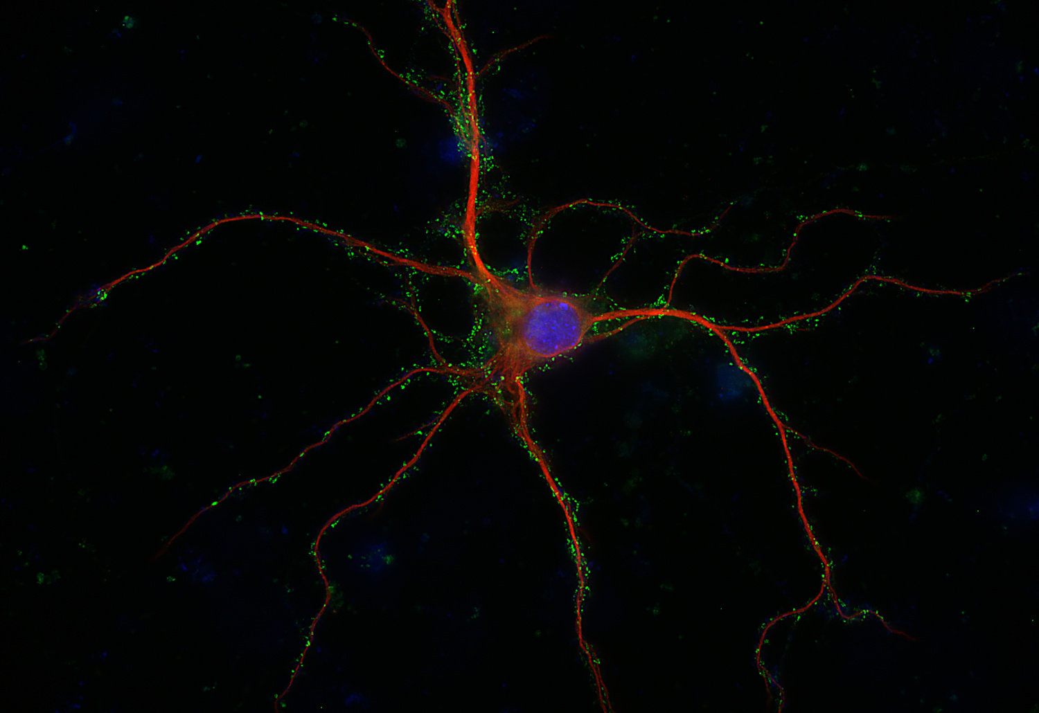
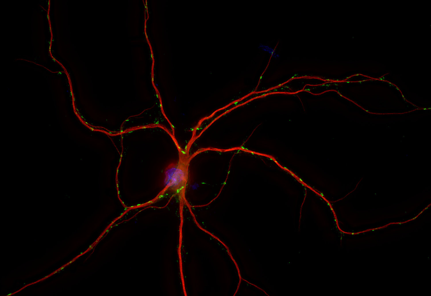
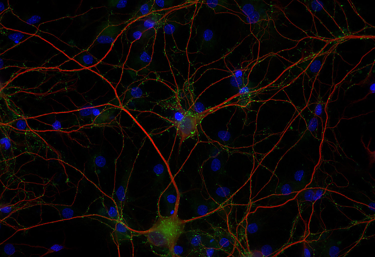
SV2s (Synaptic Vesicle Protein 2) are integral membrane glycoproteins present in synaptic vesicles. They have 12 transmembrane domains predicted by sequence analysis (1). There are three characterized isoforms, SV2 A, SV2 B and SV2 C that are similar in structure but show different expression patterns. SV2 A is expressed ubiquitously throughout the brain and plays a crucial role in modulating synaptic transmission by regulating the expression and trafficking of synaptotagmin, a key calcium sensor in neurotransmitter release (1).
SV2 B has a more restricted distribution with varying degrees of coexpression with SV2 A and is predominantly found in the cortex and hippocampus (2). SV2 C is more closely related to SV2 A but shows a very restricted expression pattern. The highest expression levels were observed in phylogenetically old brain areas like pallidum, the midbrain and the olfactory bulb (3).
SV2 expression has also been observed in other non-neuronal organs. In kidney it localizes to podocytes and is essential for the integrity of the glomerular filtration barrier (4).