Tailor-made Antibodies
and Tools for Life Science
Home | | | | | Technical Support

Synaptoporin (p38-2) antibody - 102 308

Synaptoporin s a synaptic vesicle membrane protein similar to synaptophysin1 enriched in Mossy fibers
Guinea pig monoclonal recombinant IgG
Cat. No.: 102 308
Amount: 50 µg
Price: $420.00
Cat. No. 102 308 
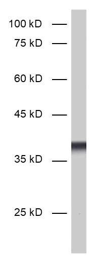
50 µg purified recombinant IgG, lyophilized. Albumin and azide were added for stabilization. For reconstitution add 50 µl H2O to get a 1mg/ml solution in PBS. Then aliquot and store at -20°C to -80°C until use.
Antibodies should be stored at +4°C when still lyophilized. Do not freeze!
Applications
 
WB: 1 : 1000 (AP staining) gallery  
IP: not tested yet
ICC: 1 : 500 gallery  
IHC: 1 : 1000 (see remarks) gallery  
IHC-P: 1 : 1000 gallery  

Western blot (WB); separation of proteins by PAGE and subsequent transfer to a membrane. Detection of target molecules is carried out with antibodies. Some antibodies require special sample preparation steps. For details, please refer to the “Remarks” section.

Immunoprecipitation (IP); Immunoisolation or pulldown of a target molecule using an antibody. For details and product specific hints, please refer to the ”Remarks” section.

Immunocytochemistry (ICC) on 4% PFA fixed cells. Immunoreactivity is usually revealed by fluorescence. Some antibodies require special fixation methods. For details, please refer to the “Remarks” section.

Immunohistochemistry (IHC) on 4% PFA perfusion fixed tissue with 24h PFA post fixation. Immunoreactivity is usually revealed by fluorescence or a chromogenic substrate. Some antibodies require special fixation methods or antigen retrieval steps. For details, please refer to the ”Remarks” section.

Immunohistochemistry (IHC-P) of formalin fixed, paraffin embedded (FFPE) tissue (some antibodies require special antigen retrieval steps, please refer to the ”Remarks” section). Immunoreactivity is usually revealed by fluorescence or a chromogenic substrate.

Clone Gp44E3
Subtype IgG2 (κ light chain)
Immunogen Synthetic peptide corresponding to residues near the carboxy terminus of rat Synaptoporin (UniProt Id: P22831)
Reactivity Reacts with: mouse (Q8BGN8), rat (P22831).
Other species not tested yet.
Matching control protein/peptide 102-1P
Remarks

The antibody is a chimeric antibody based on the monoclonal mouse antibody clone 44E3. The constant regions of the heavy and light chains have been replaced by guinea pig specific sequences. Therefore, the antibody can be used with standard anti-guinea pig secondary reagents. The antibody has been expressed in mammalian cells.
IHC: In addition to the expected strong mossy fiber staining, this antibody shows some weak unspecific staining of cell bodies in some brain regions.

Data sheet 102_308.pdf
Cat. No.: 102 308
Amount: 50 µg
Price: $420.00
Background

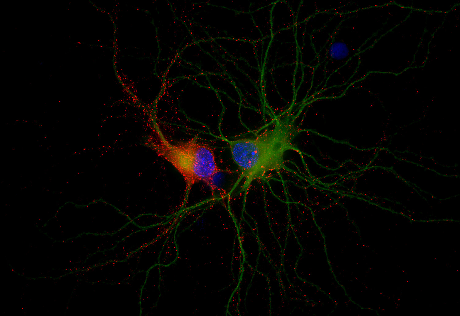
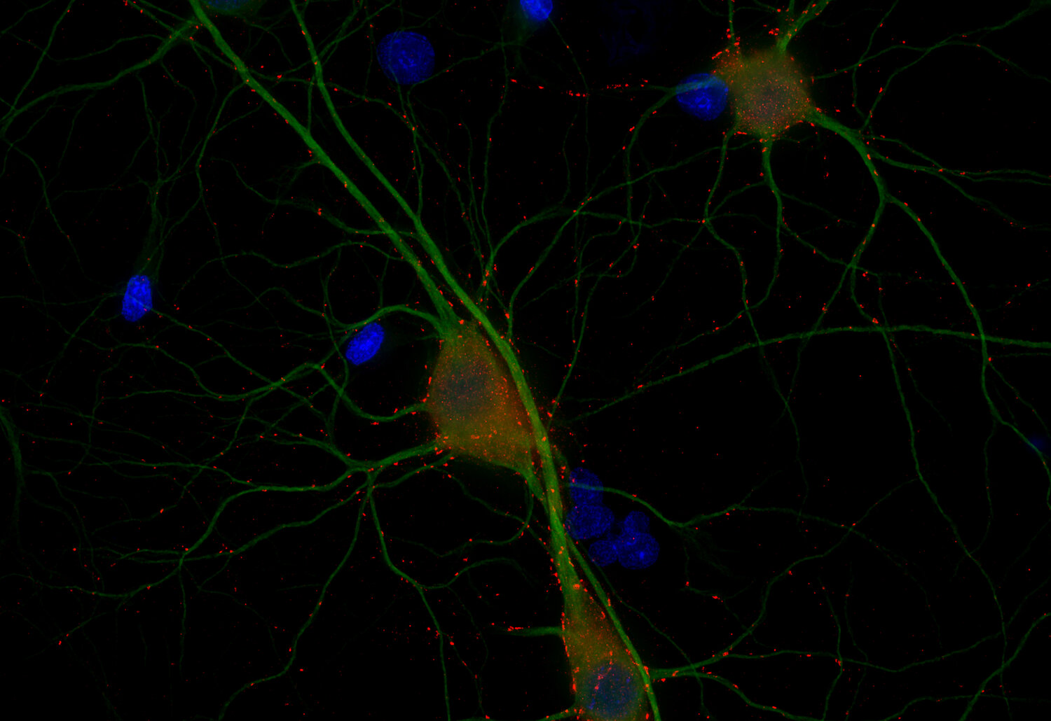
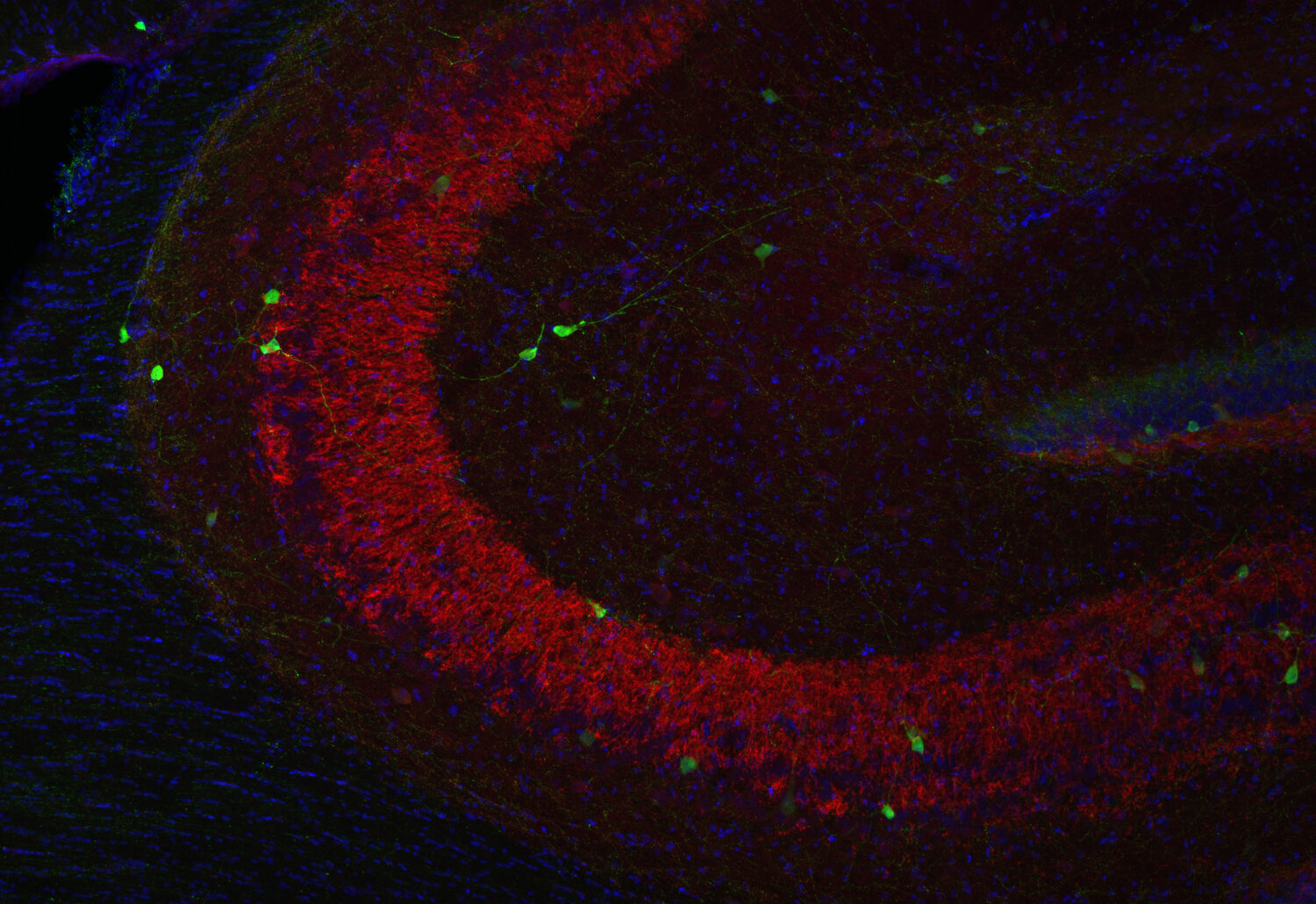
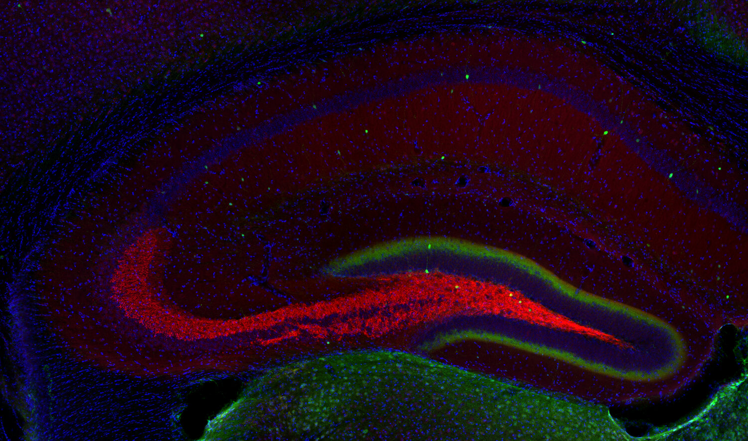
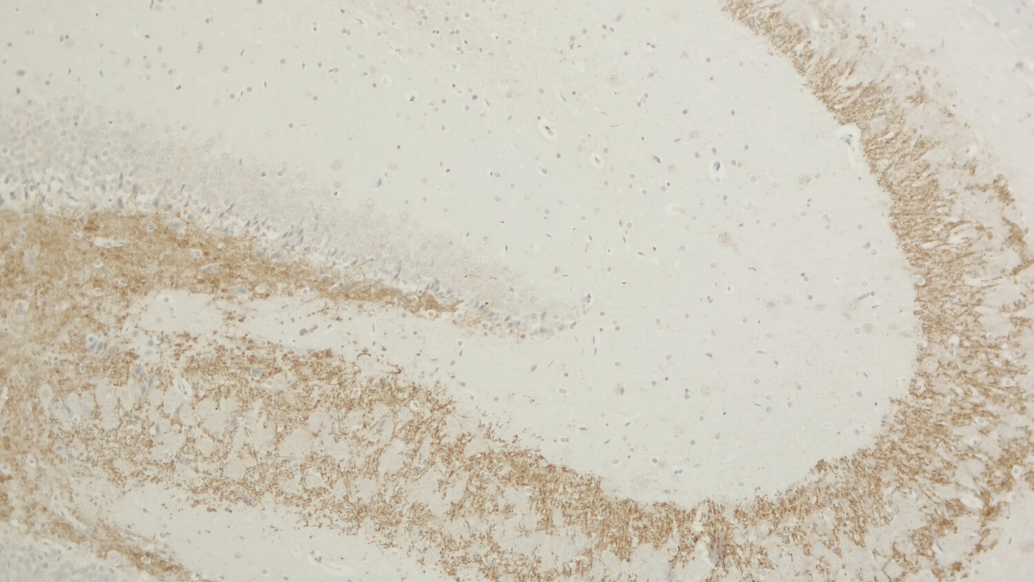
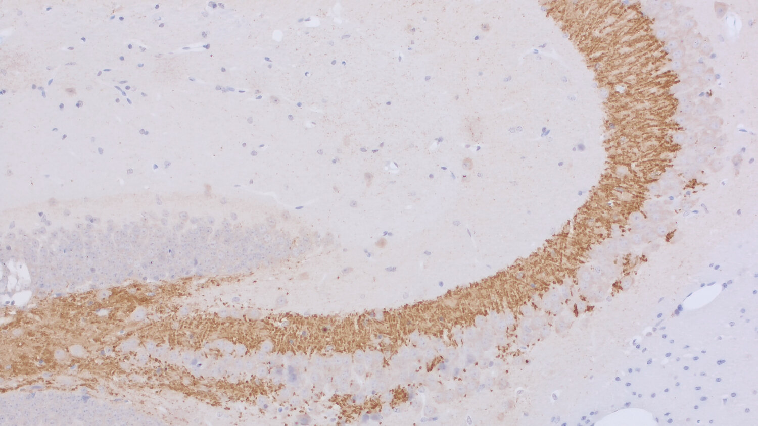
Synaptoporin, also known as synaptophysin2 and p38-2, is highly homologous to synaptophysin1 but encoded by a different gene. Like synaptopysin1, synaptoporin contains four transmembrane regions and a short cytoplasmic tail. Unlike synaptophysin1, it is not glycosylated.
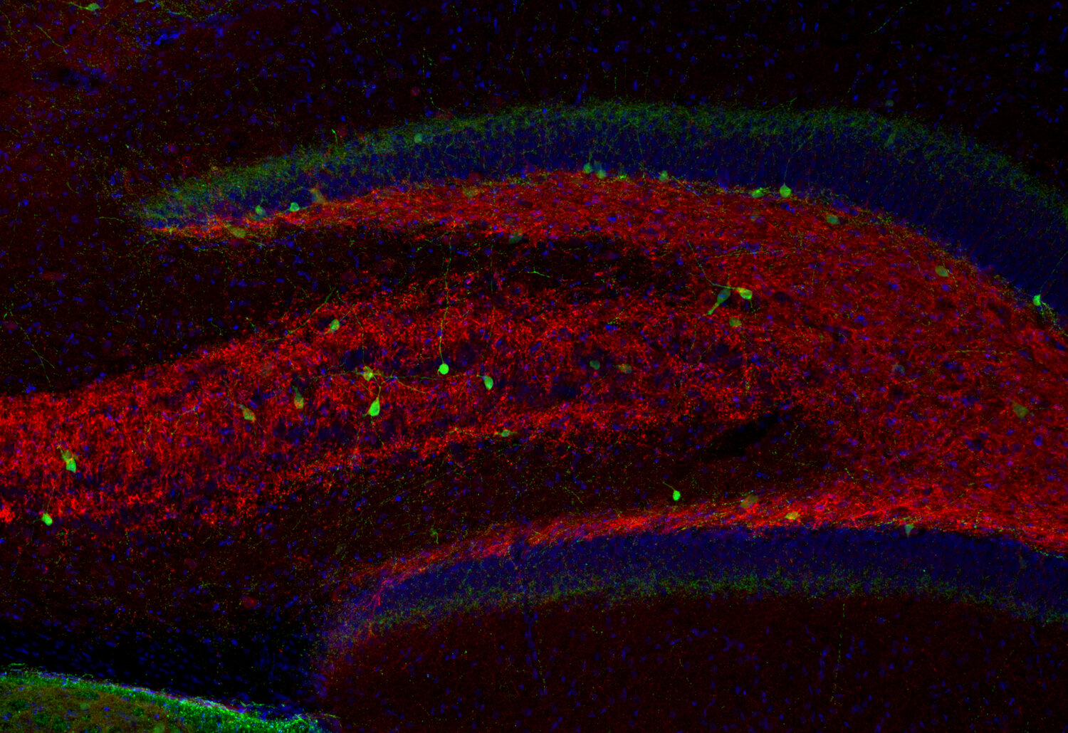
The distributions of synaptophysin1 and synaptoporin are different. Synaptophysin1 is more uniformly expressed whereas synaptoporin is particularly enriched in mossy fiber synapses in the hippocampus. It is thus an excellent marker for subsets of synapses.