Cat. No.: 160 111
Amount: 100 µg
Price:
$420.00
|
|
|
|
| Cat. No. 160 111 |
100 µg purified IgG, lyophilized. Albumin and azide were added for stabilization. For reconstitution add 100 µl H2O to get a 1mg/ml solution in PBS. Then aliquot and store at -20°C to -80°C until use. Antibodies should be stored at +4°C when still lyophilized. Do not freeze! |
| Applications | |
| Clone | SY-72G2 |
| Subtype | IgG1 (κ light chain) |
| Immunogen | Recombinant protein corresponding to the c-terminal half of human Homer1b (UniProt Id: Q86YM7-1) |
| Reactivity |
Reacts with: mouse (Q9Z2Y3), rat (Q9Z214), human (Q86YM7-1). Other species not tested yet. |
| Specificity | Specific for Homer1b and 1c; no cross-reactivity to Homer1a. |
| Remarks |
IHC: Antigen retrieval with citrate buffer pH 6 can be applied to improve the signal to noise ratio. |
| Data sheet | 160_111.pdf |
|
|


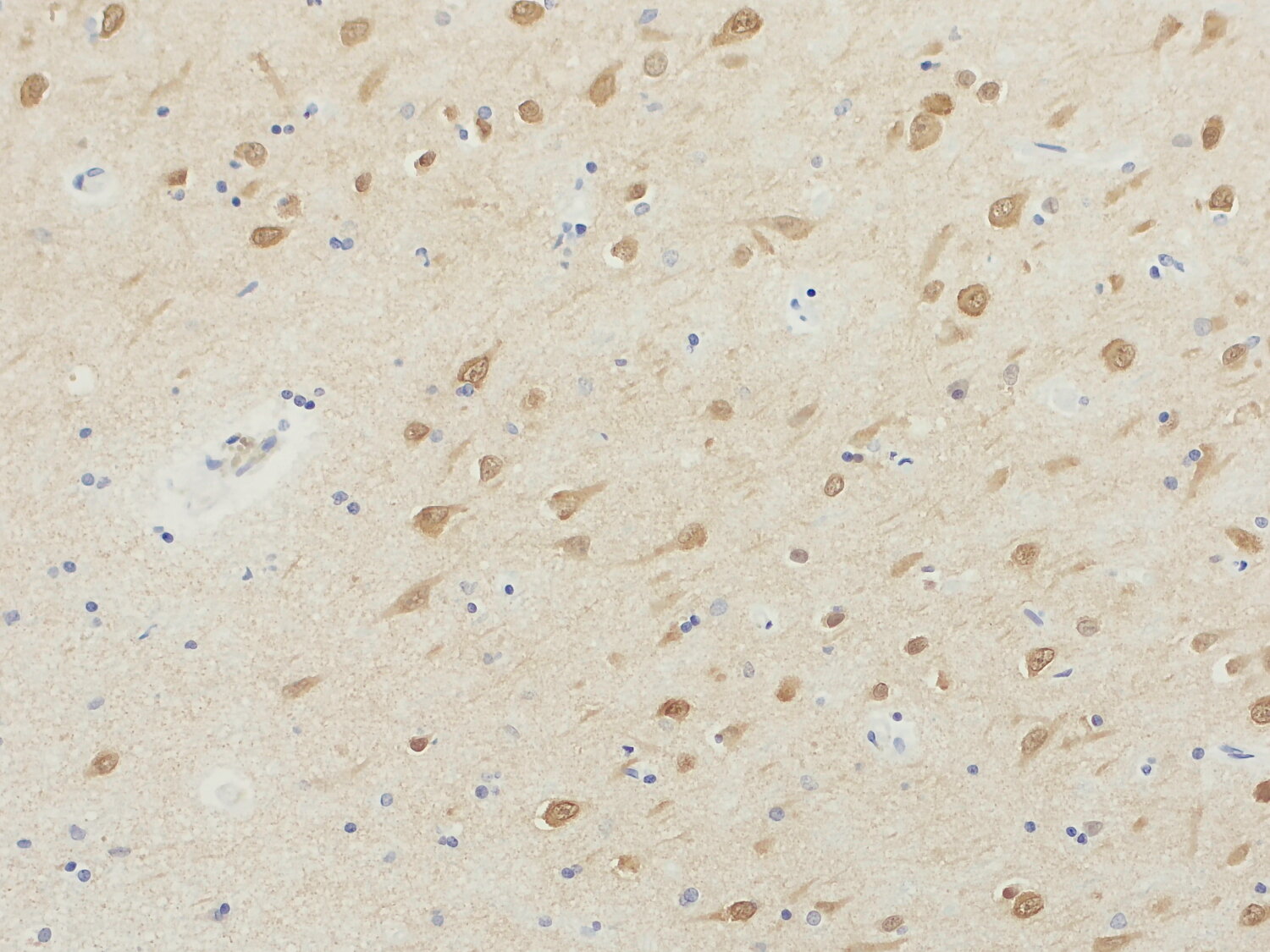
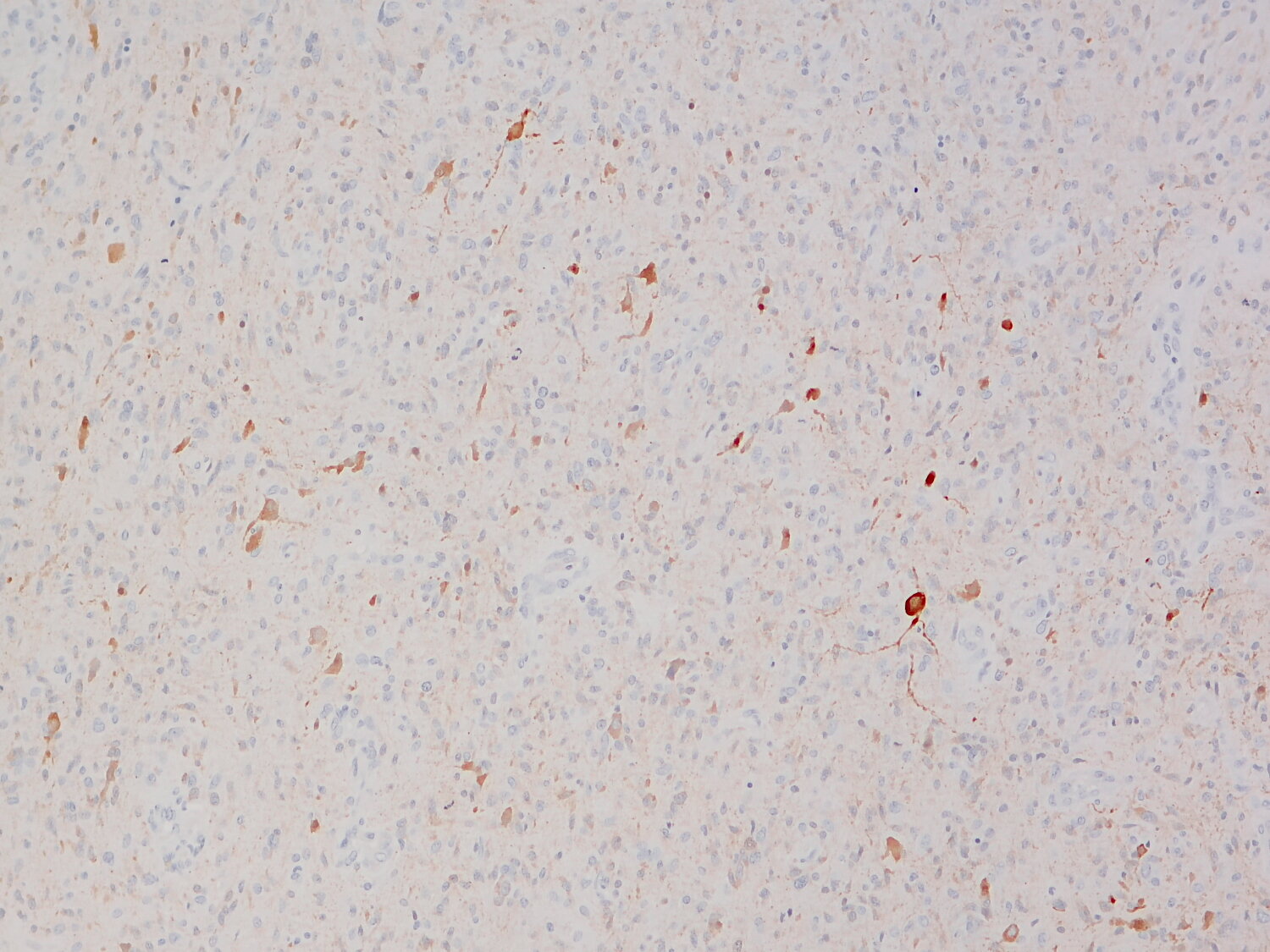
Homer is a scaffolding protein localized in the postsynaptic density (PSD) and is highly enriched at excitatory synapses. It acts as a molecular adaptor by binding to metabotropic glutamate receptors (mGluRs) (1), TRPC1 channels, Shank family proteins (2), and several other signaling molecules, organizing them into distinct clusters and thereby establishing specific signaling domains within the PSD.
By cross-linking these proteins, Homer plays a crucial role in structural and functional organization of the PSD, contributing to the maturation of dendritic spines and the regulation of synaptic plasticity. Homer and Shank, in particular, form a mesh-like matrix that serves as a platform for assembly of other PSD proteins (3).
There are three main Homer isoforms—Homer1, Homer2, and Homer3—each of which is subject to alternative splicing, producing multiple splice variants such as a, b, c, and d. These variants can have distinct functional properties, and their dynamic redistribution at synapses is involved in remodeling the PSD in response to neuronal activity (4).
Emerging evidence suggests broader roles for Homer1b/c beyond synaptic scaffolding, including in non-neuronal contexts, although their specific involvement in cancer remains unclear (5).